Magnetic Resonance Imaging
Superman may have had X-ray vision but even he couldn’t see as much as we can today by using Magnetic Resonance Imaging or a MRI scan.
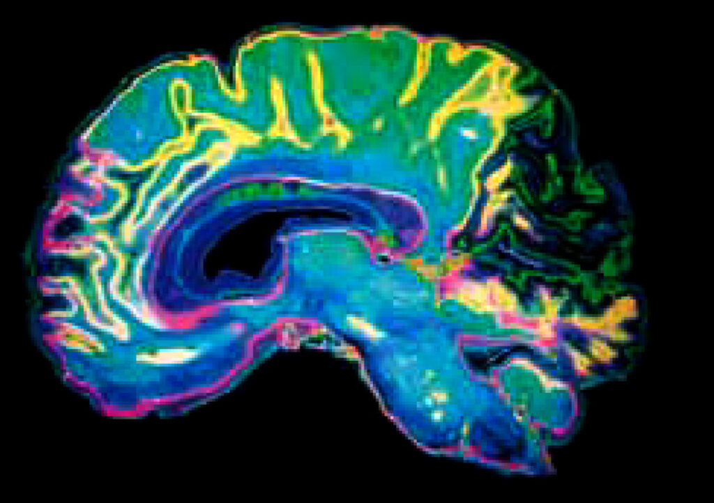
MRI is a safe and painless test that uses a strong magnetic field and radio waves to create pictures, of tissues, organs and other structures inside your body, meaning that there is no exposure to x-rays or any other damaging forms of radiation. These images are comparable to an anatomy drawing in a textbook.
Why do we need this innovation?
An MRI scan can create clear pictures of most parts of the body. It is useful for all sorts of reasons where other tests (such as X-rays) do not give enough information or are not clear enough, is commonly used to get detailed pictures of the brain and spinal cord, or to detect abnormalities and tumours. Even torn ligaments around joints can be detected by an MRI scan. It is being used more and more following sports injuries, with many Premiership footballers and Olympic athletes undergoing MRI scans before being declared sports fi t.
What does an MRI scan involve?
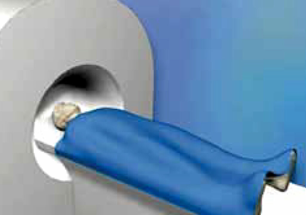
The MRI scanner is like a tunnel about 1.5 metres long surrounded by a large circular magnet. You lie on a couch which then slides into the scanner. A ‘receiving device’, like an aerial, is placed behind, or around, the part of the body being examined. This detects the tiny radio signals emitted from your body. When each ‘picture’ is being taken you need to keep still for a few minutes, otherwise the scan picture may be blurred.
The scan itself is painless and can take 15-60 minutes in total, although no individual scan is longer than 10 minutes. It may be a little uncomfortable lying still on the couch for this time. In some cases, an injection of a special contrast dye is given into the bloodstream via a vein on the arm. This helps to give clearer pictures of certain tissues or organs being examined.
Attack of the Facts
Fact 1
MRI scanning allows the doctor to view what is going on inside the body without the use of x-rays. MRI allows them to discover understand what’s not working without the need for exploratory surgery: therefore, reducing the need for unnecessary operations.
Fact 2
The technology is constantly evolving, and the next generation scanners will dramatically reduce the time a patient spends in the scanner. It will be easier to image the whole body rather than just small areas of it.
Fact 3
MRI scans are better than X-rays at showing soft tissue or inflammation, for detailing blood vessels and creating cross-sectional pictures in any plane or direction. The scanners can also tell us about the body’s function not just anatomy.
Fact 4
The scientific principles behind MRI were discovered in 1946, but it was not until the 1970s that the technology became available to use these principles. The first commercial MRI scanner was installed in Manchester in 1983.