Summary

Flow cytometry in the cellular laboratory is largely divided into two areas; primary immunodeficiency and leukaemia diagnosis. For the purposes of diagnosing immunodeficiency, blood cells are checked for the presence of intact surface receptors that mediate effective immune responses. Flow cytometry allows scientists to label specific surface receptors on different populations of blood cells so that this functionality can be investigated. In leukemia a mutation in genetic expression leads to the uncontrolled proliferation of a single lineage of immune system cells. Flow cytometry enables scientists to establish what particular type of leukaemia a patient has by checking their blood cells for different sets of surface molecules that typify different leukemias. It is important to characterise different leukemias because the type can have a significant effect on patient prognosis and treatment.
Science
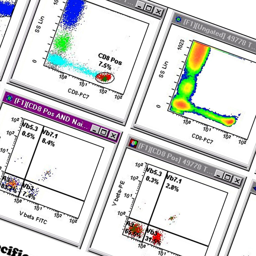
Individual populations of immune system cells with specific roles and at different stages of maturation have different groups of molecules on their surface, this characteristic is useful for quantifying the different populations as we can identify them based on these specific markers. As with many other techniques used in the immunology laboratory, commercially available antibodies that have specificity for single unique surface molecules are mixed with patient blood so that they can bind to their targets. These antibody labels are joined to dyes (fluorochromes) that emit light at specific wavelengths when excited by light at a lower wavelength. Several different colours attached to antibodies with different specificities can be used in the same sample. Cells labelled with the different antibodies are rapidly passed in single file through a laser beam that excites the dye molecules. The light that is emitted at different at different wavelengths is detected through a series of finely aligned detectors. In addition the way that the cell itself interacts with the laser beam is recorded and these measurement are extrapolated to determine how big and granular they are. The results are represented graphically and by using this data every cell is recorded as having a specific signature based on size, granularity, and abundance of surface markers.
In leukemia a single type of cell at a specific stage of development undergoes rapid proliferation so that it represents the majority of cell types, this in turn results in a reduction of all other functioning cells. Using flow cytometry this population can be determined as a single population of cells with a discrete set of surface marker characteristics that provides information about what type of leukemia it represents.
Clinical

Leukaemia is a disease of the blood or bone marrow resulting from the cancerous proliferation of cells of the immune system, caused by a mutation in a single stem cell. Patients can present with a variety of symptoms, some of the more common include bone pain, excessive bruising, anemia and uncontrolled infections. Chronic leukaemias will cause death if untreated in months or years however acute leukaemias will can cause death in weeks or months. A multi-disciplinary approach, utilising flow cytometry in conjunction with clinical history, morphology, full blood counts and cytogenetics to diagnose specific types of leukemia is routinely employed. This protocol is important as often a subtly different diagnosis can have profound effects on patient prognosis.