Summary
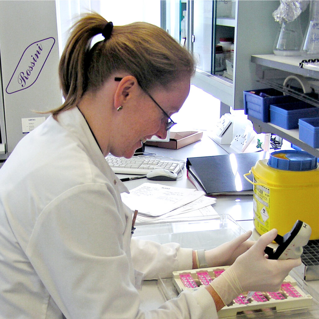
The technique of Immunofluoresence involves an ultra violet light microscope that can visualise fluorescently labelled autoantibodies in patient serum when applied to tissue sections on microscope slides. The results from this can be used to diagnose and monitor autoimmune diseases such as SLE, Diabetes, and Myasthenia Gravis.
Science
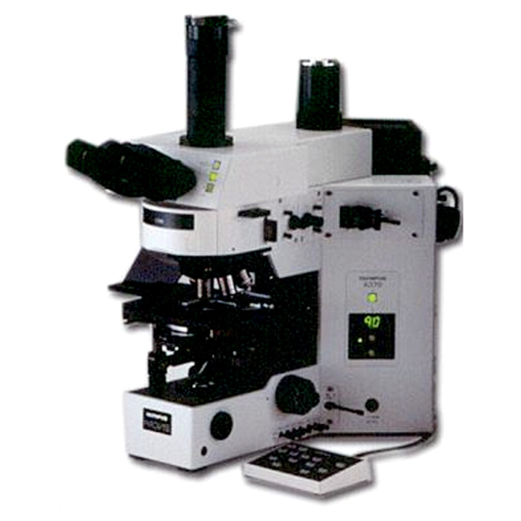
In patients with a suspected autoimmune disease it is often useful to identify the presence of autoantibodies that are directed against specific tissues. These autoantibodies are often not the cause of the disease but can be useful markers of disease association with varying degrees of specificity. Patient serum is separated from the red blood cells and then applied directly onto a specific tissue section (substrate) mounted a microscope slide, the type of tissue substrate is chosen as having relevance to the suspected disease. After a period of incubation, the serum is washed off leaving any autoantibodies bound to specific antigens of the substrate. The next step is to add a fluorescently labelled antibody that will bind to all human antibodies, therefore binding to and fluorescently identifying any of the patient’s antibodies that have bound to the tissue. After a final incubation any excess conjugate is washed off and the sample exposed to UV light under a specialised microscope. The fluorescent label absorbs UV light, becomes excited and emits light at a different wavelength (green light) (Physics curriculum) so areas on the substrate where patient antibodies have bound can be visualised. Patient samples are compared with negative and positive controls to identify where significant autoantibody presence could indicate a specific disease.
Clinical

Several diseases can be diagnosed with the aid of indirect immunofluoresence techniques on different tissue substrates.
- A diagnosis of Systemic Lupus Erythematosus (SLE) can be aided by identifying autoantibodies directed against DNA in cell nuclei of human epithelial cells. This test might be indicated in a patient who presented with any one of a number of systemic symptoms including rashes, kidney damage, vasculitis, photosensitivity, vasculitis and neurological problems (curriculum)
- Diabetes can be diagnosed by identifying autoantibodies against the pancreatic islet cells that produce insulin in healthy people. A diabetic patient could present to their doctor with fatigue, frequent urination and increased thirst.
- A doctor may suspect Primary Billiary Chirrosis in an individual with jaundice, itchy skin and general fatigue. To assist this diagnosis he or she may ask the immunology laboratory to look for patient antibodies against mitochondrial anitgens found in most cells. This would be investigated using liver and kidney tissue sections.
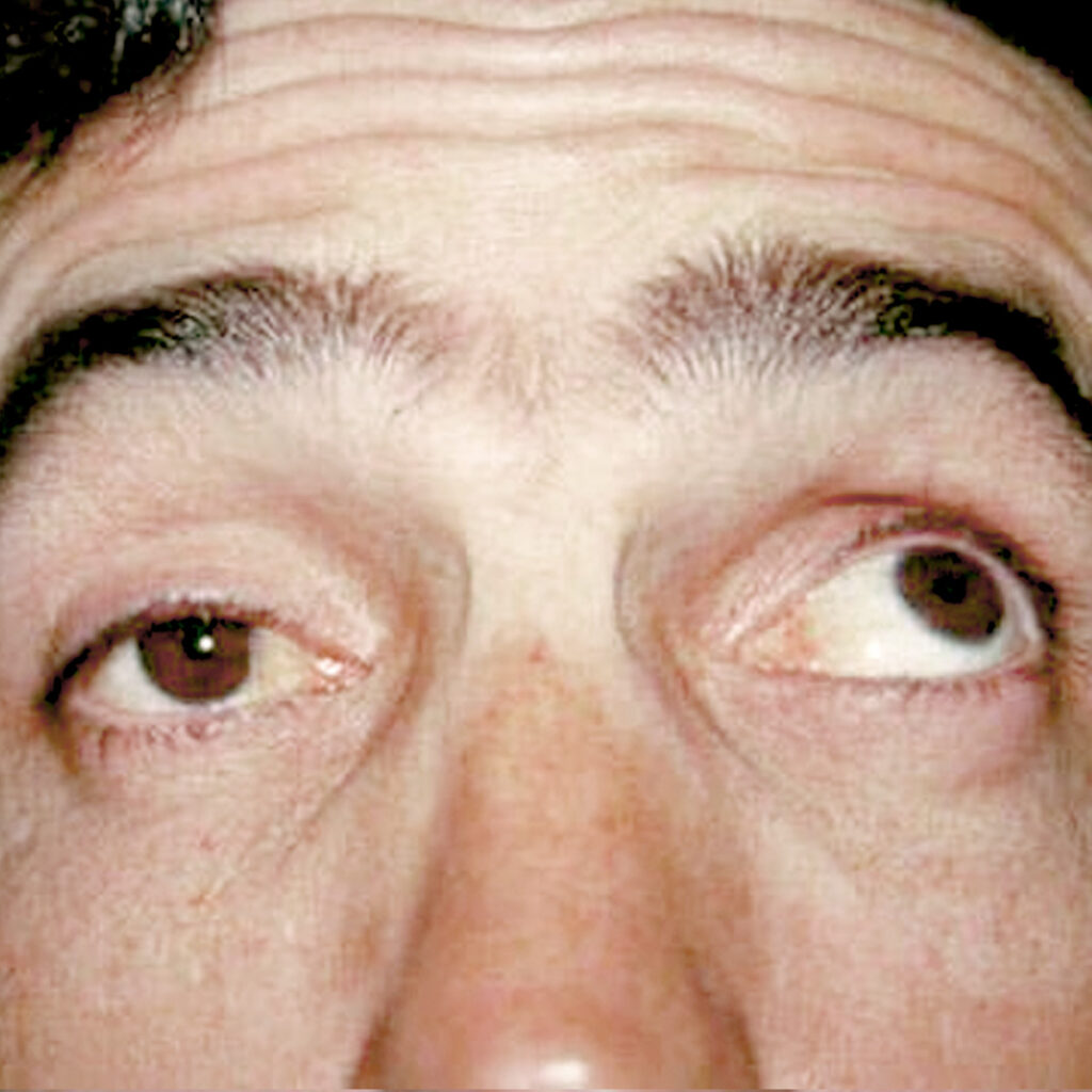
- Myasthinia Gravis is a rare condition where the patient has progressive muscle weakness which can lead to difficulty swallowing and problems with eye coordination. Sections of striated muscle fibre are used here to identify specific autoantibodies.